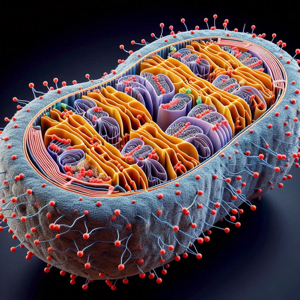
Researchers are now able to observe the dynamic processes of respiration at the atomic level in a natural membrane environment for the first time thanks to innovative high-resolution microscopy technologies. The new method may make it easier for scientists to comprehend what’s going on within sick cells’ mitochondria and other organelles and may also help them find new, more targeted therapeutic targets.
The findings were published in the journal Nature in May.
By combining two microscopy techniques single-particle analysis and cryo-electron tomography, or cryo-ET researchers created a unique imaging method for visualizing mitochondria from animal models. In order to address obstacles to high-resolution cryo-electron microscopy (cryo-EM) interpretation of pictures from the extremely congested environment, they also created novel computational techniques.
With the use of this technique, the team was able to observe protein structures in the organelle at a resolution never before possible. They also looked at how the architecture of these proteins inside the mitochondrial membrane changed when illness was induced in these mice. Their research opens the door to an atomic-level understanding of the molecular makeup of cells.
Previously, our technology was not able to reveal how interactions occur,
But in this case, we see the atoms, we see the structures of molecules, and we see how everything interacts, giving us an entirely new understanding of the mechanisms in the cellular environment.
Jack Zhang, PhD
Structural biologists are now constrained by the resolution or target scales that can be reached by the existing microscopy methods.
We haven’t even been able to distinguish protein subunits [using conventional approaches], let alone atomic details in cellular environments.
Jack Zhang, PhD
Combining the ideas of cryo-EM and cryo-ET technologies for single-particle analysis with newly developed image processing methodologies to analyse high-resolution cellular cryo-EM pictures at the cryo-ET scale allowed the Yale team to achieve their remarkable achievement.
Single-particle analysis is great for imaging individual molecules, but not for cellular structures,
Cryo-electron tomography is a perfect tool to study cellular structures, but its resolution is typically limited to several nanometers.
Jack Zhang, PhD
By creating a method to photograph cryo-ET targets in single-particle mode and using cryo-ET reconstructions as first references for single-particle alignment and classification later on, they surmounted this technological difficulty.
Also Read| Cancer can be blocked by Statin therapy, which can block the inflammatory protein
Conventional single-particle cryo-EM has another drawback in that technique usually necessitates protein purification and isolation prior to freezing and mounting the molecules onto cryo-EM grids, which removes them from their natural habitat. This mechanism breaks down physiological responses as well as weakly formed higher-order biological complexes. This is especially significant for membrane proteins, as the native membrane environment which is essential to their regular functions would be totally destroyed by detergent-purified samples.
Previously, our technology was not able to reveal how interactions occur. But in this case, we see the atoms, we see the structures of molecules, and we see how everything interacts.
Jack Zhang, PhD
Researchers can now view molecular structures with an unprecedented level of detail thanks to the team’s innovative algorithms that bridged the gap between single-particle resolution and cryo-ET scale images created by combining these microscopy approaches with new computational techniques. Interestingly, their method made it possible for scientists to observe membrane protein structures inside of native mitochondria for the first time, as well as the proteins’ normal construction and activity across the whole organelle.
The team was able to see the atomic-level structures of the many reactive intermediates as they work within mitochondria since all of these supercomplexes were in their natural environment, which is different from isolated proteins.
Also Read| A father’s diet can influence his children’s health through sperm
Using a novel classification strategy, we see how proteins are reacting to their environment and captured different intermediate states,
Conventional approaches only provide location information of molecules or their overall shapes at low resolution in cellular environments. This new approach has substantial pharmaceutical and clinical implications. “It can help us study how molecules react to certain drugs in their native cells.
Jack Zhang, PhD
Additionally, this microscopy method may be revolutionary in aiding researchers in comprehending the fundamental causes of a variety of ailments. The Yale researchers produced animal heart models of different stages of myocardial ischemia, a cardiovascular disease in which a blockage prevents blood flow to the heart, in order to investigate this further.
They discovered that in addition to their original membranes, the various phases of the illness significantly altered the conformational distribution and shape of the supercomplexes.
Also Read| Scientists discovered a protein that has a key role in human blood stem cell self-renewal
With this unique insight into the workings of both healthy and sick cells, the Yale team’s method may potentially lead to the creation of extremely precise treatments that specifically target these newly discovered molecular pathways or structures. This is especially true for membrane proteins, which play important roles in physiological and pathological processes such as signal transduction, pathogen-host interactions, cell-cell communication, and high specificity and selectivity. They are also highly accessible to drugs, have a wide range of target classes, and have significant therapeutic potential.
Source: Yale School of Medicine News
Journal Reference: Zheng, Wan, et al. “High-resolution in Situ Structures of Mammalian Respiratory Supercomplexes.” Nature, 2024, pp. 1-8, https://doi.org/10.1038/s41586-024-07488-9.
Last Modified: